Thanks to bone-forming and bone-replacing preparations and collagen membranes, it is possible to perform procedures to rebuild atrophied tooth-bearing structures - regeneration of bone damaged by periodontitis, bone augmentation prior to implant placement, reconstruction of the soft tissues surrounding the tooth. The preparations used in our clinic are the best studied on the market and have the largest database of clinical studies, in a word, they are absolutely safe.
If you want to see how we use biomaterials - watch the video!
If you want to understand what bone regeneration is all about - watch the video!
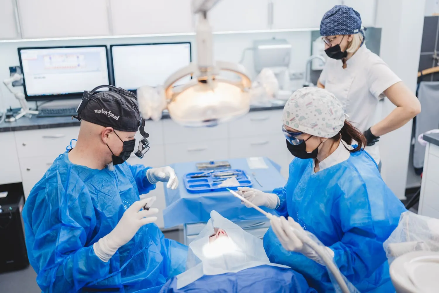
Use of biomaterials:
Periodontopathies - This damage is caused by bacteria in dental plaque. Their deposits lead to the destruction of the tissues that support the teeth; the supporting tissue of the tooth atrophies and the ligamentous apparatus of the tooth is destroyed together with the surrounding bone, leading to tooth loss.
Apical lesions - This effect is caused by inflammation at the apex of the root. This type of inflammation leads to destruction of the bone structure.
Extractions - The empty socket after a tooth extraction should be filled with bone-forming granules to prevent alveolar atrophy.
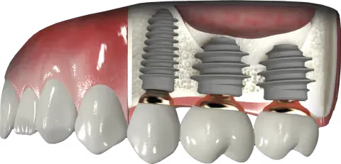
Implantation
If the patient's own bone volume is not sufficient to insert the implant.
If the insufficient bone supply does not cover the entire implant.
If the alveolar process is too narrow.
If the height of the alveolar process is insufficient.
Sinus lift - Sinus floor lift
Indications This procedure considerably extends the use of implants in patients with insufficient bone in the lateral jaw sections (a very common situation). It makes it possible to avoid wearing removable prostheses. The regenerated bone is an excellent "support" for the implants, so that natural-looking crowns and bridges can be used. The patient has the impression that they have their "own" teeth.
Course of treatment - (3-phase version)
The period of complete bone regeneration after the sinus lift is 6–9 months (unnoticed by the patient). This is followed by implant placement. After a period of 3 to 6 months following implantation, the prosthetic work (crowns, bridges, dentures) is inserted.
Treatment procedure - (2-step version)
If the anatomical conditions (thickness of the maxillary sinus floor) allow, the implant procedure is carried out at the same time when bone regeneration is required. In this case, the healing phase (6-9 months) is followed by insertion of the prosthetic work. This saves 3–6 months of time.
Diagnostics
Before performing a sinus lift, we routinely carry out a CT scan to familiarize ourselves with the exact anatomy of the maxillary sinus. This allows us to determine the degree of difficulty of the procedure, rule out possible inflammation, and decide whether immediate implant placement is possible.
Sinus lift surgery
Description - the procedure is performed under local anesthesia and is completely painless.
1. Creation of an access "window".
Enables access to the maxillary sinus, the "floor" of which is too narrow to insert implants.
2. Elevation of the mucosa lining the maxillary sinus
Creates space for future/regenerated bone.
3. Filling the space created with bone substitute material
Insertion of bone-forming material (bone will form in its place after 9–12 months) into the maxillary sinus.
4. Closure
Sealing of the access window with a barrier membrane and suturing of the mucosa.
5. Implantation
If the anatomical conditions allow, the implants can be inserted during the operation, which shortens the duration of the entire treatment.
6. Integration of the implants.
After a period of around 6 months, the implants are fully integrated into the reconstructed jawbone.
After the treatment
There is a healing phase after the procedure. The first phase lasts 2–3 days, during which swelling and slight pain occur. The patient is given appropriate painkillers, antibacterial rinses - rest is recommended (no sport) and a semi-fluid, cool diet. The next phase lasts about 10 days - the swelling disappears, the stitches are removed, the patient begins to function normally, the diet is still soft.
Contraindications
Severe systemic diseases, unstabilized high blood pressure or diabetes, radiotherapy, severe anatomical conditions. All conditions, the treatment method and prognosis are discussed in detail at the consultation appointment with the implantologist after a thorough examination of the patient.